Kyla
The Cardiovascular System
What is the Cardiovascular System?
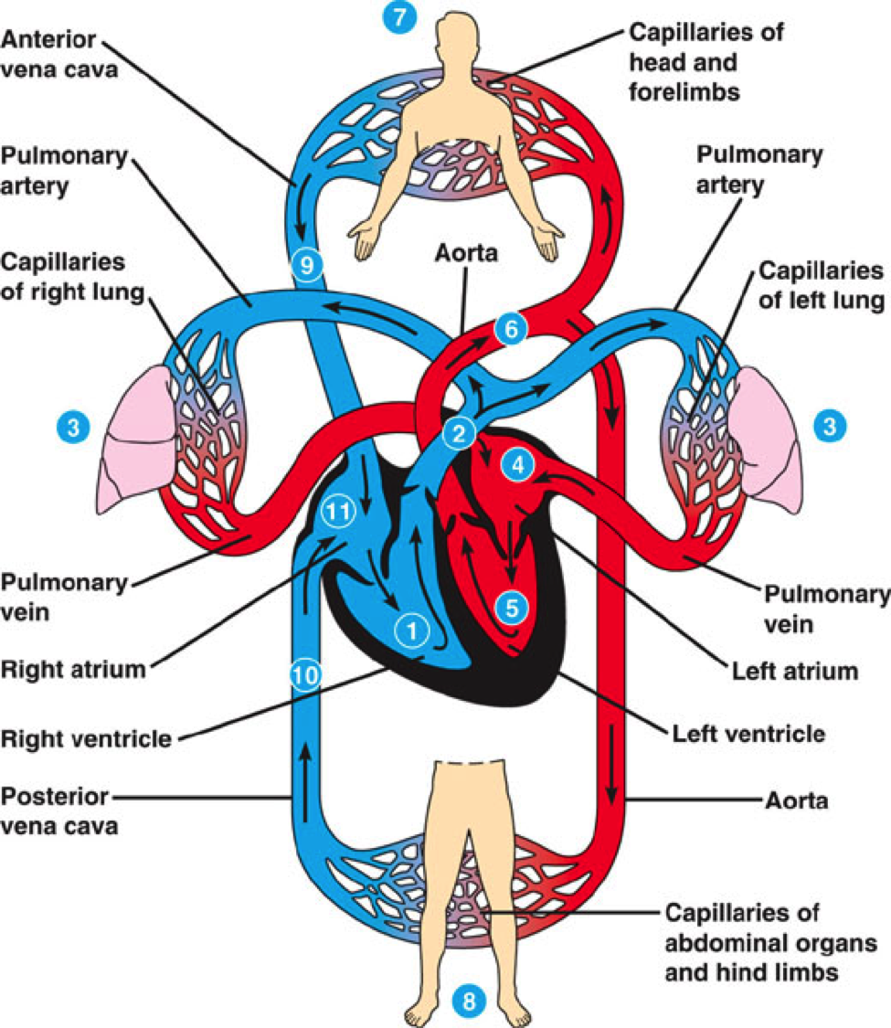
The cardiovascular system delivers nutrients and oxygen to all cells in the body. It consists of the heart and the blood vessels running through the entire body. The arteries carry blood away from the heart then the veins carry it back to the heart. The cardiovascular system consist of 4 major organs the heart, veins, arteries, and capillaries.
How does the cardiovascular system work?
The heart, blood, and blood vessels work together to service the cells of the body. Using the network of arteries, veins and capillaries, blood carries carbon dioxide to the lungs and picks up oxygen. From the small intestine, the blood gathers nutrients and delivers it to every cell.
Organs in the Cardiovascular System
Heart
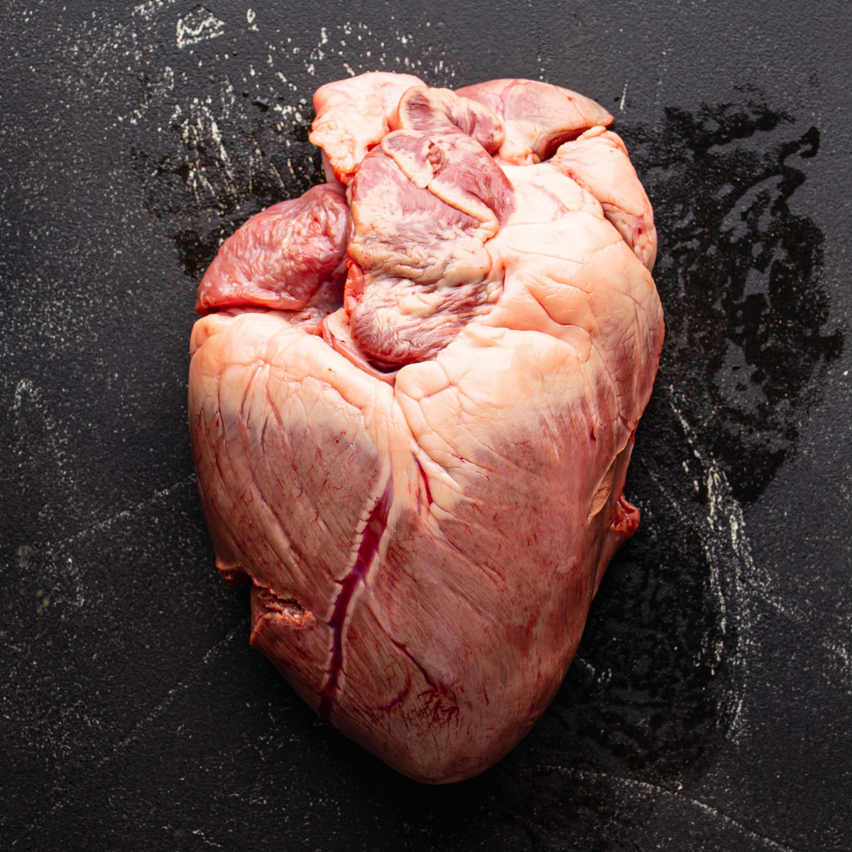
The heart is a fist-sized organ that pumps blood throughout your body. It's the primary organ of your circulatory system. Your heart contains four main sections (chambers) made of muscle and powered by electrical impulses. Your hearts main function is to move blood throughout your body. Your heart controls the rhythm. Your heart works with other body systems to control your heart rate and other body functions. The primary systems are the endocrine and the nervous system. Your nervous system helps control your heart rate. It sends signals that tell your heart to beat slower during rest and faster during stress. Your endocrine system sends out hormones. These hormones tell your blood vessels to relax, which affects your blood pressure. Hormones from your thyroid gland can also tell your heart to beat faster or slower. Your heart is generally located on the left side because your heart's left ventricle has to be bigger because it pumps oxygen rich blood around the whole body.
Veins
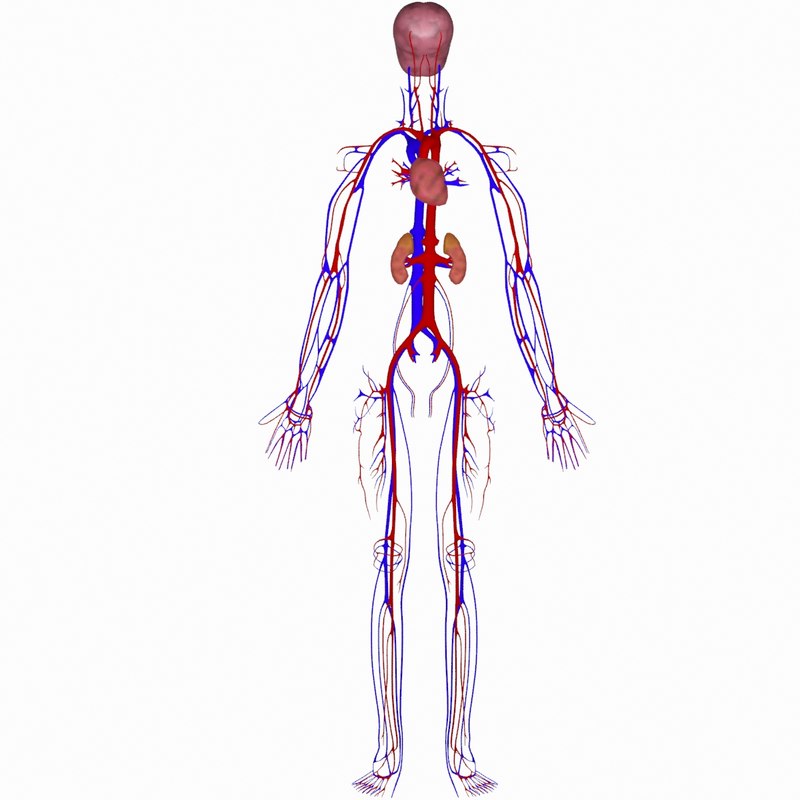
Veins are blood vessels located throughout your body that collect oxygen-poor blood (with an exception to your pulmonary veins) and return it to your heart. They work together with other blood vessels and your heart to keep your blood moving. Many people think veins are blue because they look blue through our skin. However, that’s just a trick that our eyes play on us. Your veins are actually full of dark red blood therefore, they are dark red. Your veins look blue because of the way light rays get absorbed into your skin. Blood is always red both in your veins and arteries never blue.
Arteries
Arteries are blood vessels that distribute oxygen-rich blood to your entire body. These tube-like vessels and the muscles inside them ensure your organs and tissues have the oxygen and nutrients they need to function. You have two types of arteries that have slightly different functions. You have your elastic artery which gets your blood from your heart before passing it on to other arteries. You also have your muscular arteries which gets blood to your tissues. Arteries get messages from your central nervous system to tighten or open up. This affects your blood pressure, or the force of your blood moving through your arteries. Arteries help keep your blood pressure steady. They also control blood flow. They do both by tightening or loosening their muscle walls.
Capillaries
Capillaries are delicate blood vessels that exist throughout your body. They transport blood, nutrients and oxygen to cells in your organs and body systems. Capillaries are the smallest blood vessels in your vascular system. Capillaries have three different shapes. Continuous fenestrated capillaries have small openings or fenestrae that enable the rapid exchange of substances. This type of capillary is in your kidneys, small intestine and endocrine glands. Continuous nonfenestrated capillaries have a lining through which only small molecules can pass. This type of capillary exists in the nervous system as well as fat and muscle tissue. Also, the sinusoidal capillaries have small fenestrae that allow certain substances to pass through. This type of capillary is in your liver and spleen.
Cardiovascular System Planes and Anatomical Directions
Cardiovascular in a Transverse Plane
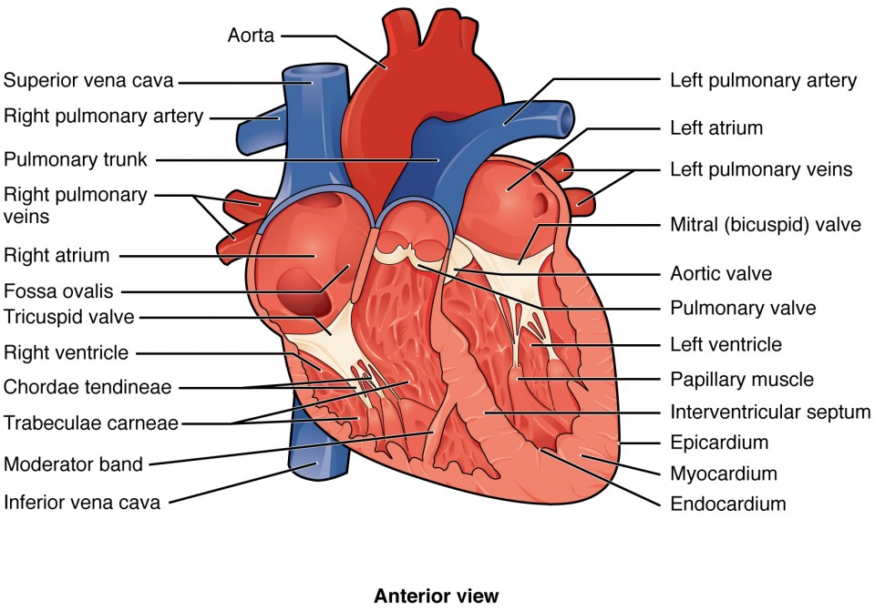
The transverse plane is an anatomical plane that divides the body into superior and inferior sections. It is a horizontal dissection across the y-axis. In a transverse plane the anatomical directions are posterior and anterior. Posterior means the back. Anterior is the opposite of posterior therefore anterior means front. A transverse plane has the anatomical directions lateral meaning to the side of and medial meaning middle. For example, in the diagram the tricuspid is lateral to the aortic valve.

Cardiovascular in Sagittal Plane
A sagittal plane is a vertical plane which passes through the body along the z-axis. The anatomical directions are posterior, anterior, inferior which means below, and superior which means above. For example, the Bicuspid valve is inferior to the aorta.
Cardiovascular in a Frontal Plane
A frontal plane is a vertical dissection running along the x-axis. The anatomical directions for a frontal plane include medial, lateral, inferior, superior, contralateral, and ipsilateral. Contralateral is on opposite sides of the body. Where as, ipsilateral is on the same side of the body. For example, the right atrium is contralateral to the left atrium.
Cardiovascular Cavities
Cavities
There are two major cavities called the dorsal/posterior cavities and ventral/anterior cavities. In the dorsal/posterior cavity we have two subdivided cavities called the spinal cavity and cranial cavity. In the ventral/anterior cavity we have three subdivided cavities such as the thoracic cavity, abdominal cavity, and pelvic cavity.
Spinal Cavity
The spinal cavity is an anatomical space formed by the vertebral column that stores an integral portion of the nervous system. In this cavity we have our dorsal spinal vein. The dorsal spinal vein receives blood from the dorsal horns and dorsal funiculi. Lateral funiculi drain directly into spinal branches of the vertebral, intercostals, lumbar and sacral veins. The ventral spinal vein runs throughout the ventral median fissure and mainly receives blood from the ventral horns.

Cranial Cavity
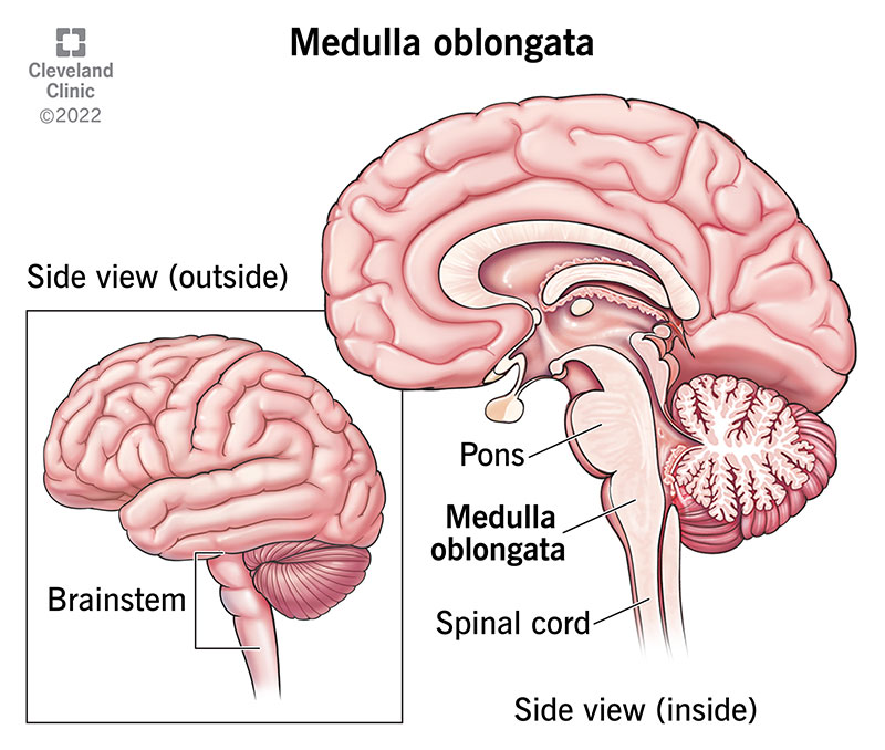
The cranial cavity is the space formed inside the skull. The cardiovascular center is a part of the human brain found in the medulla oblongata. The medulla oblongata is responsible for regulation of cardiac output. The medulla oblongata is the connection between the brainstem and the spinal cord, carrying multiple important functional centers. It is comprised of the cardiovascular-respiratory regulation system, descending motor tracts, ascending sensory tracts, and origin of cranial nerves
Thoracic Cavity
Your thoracic cavity is a space in your chest that contains organs, blood vessels, nerves and other important body structures. It's divided into three main parts, the right pleural cavity, the left pleural cavity as well as the mediastinum. The five organs in your thoracic cavity are your heart, lungs, esophagus, trachea and thymus. The only one of these organs that is part of the cardiovascular system is the heart. The heart is responsible for sending oxygen and nutrients to all parts of your body, and carries away unwanted carbon dioxide and waste products.

Abdominal Cavity
The abdominal cavity is the space between the abdominal wall and the spine. The abdominal cavity contains a number of crucial organs including the lower part of the esophagus, the stomach, small intestine, colon, rectum, liver, gallbladder, pancreas, spleen, kidneys, and bladder. The abdominal cavity also has many blood vessels. The major arteries of the abdominal wall are the superior epigastric, inferior epigastric, musculophrenic, subcostal, and posterior intercostal arteries, deep circumflex iliac artery, superficial circumflex iliac artery, and superficial epigastric artery. These vessels help supply and drain the posterior abdominal wall, and the abdominal viscera.

Pelvic Cavity
The pelvic cavity is a bowl-like structure that sits below the abdominal cavity. The pelvic floor supports the urinary bladder, uterus in females, pelvic colon, rectum. The pelvis floor also functions as a bladder and anal sphincter through tonic contractions. In the pelvic cavity we have the inferior vena cava. The inferior vena cava is responsible for venous drainage of the abdomen. The blood from the portal vein passes through the liver and finally drains into the inferior vena cava. The inferior vena cava forms at the level of the fifth lumbar vertebra by the joining of left with the right common iliac veins.

Tissues
What are tissues?
Tissue is a group of cells that have similar structure and that function together as a unit. There are different types of tissues that serve different purposes. We have four main types of tissues including the Epithelial tissues, connective tissue, muscle tissue, and nervous tissue.

Epithelial Tissue
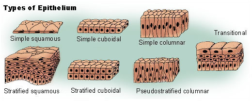
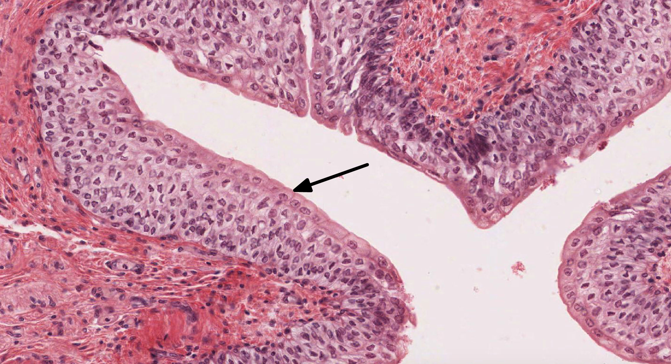
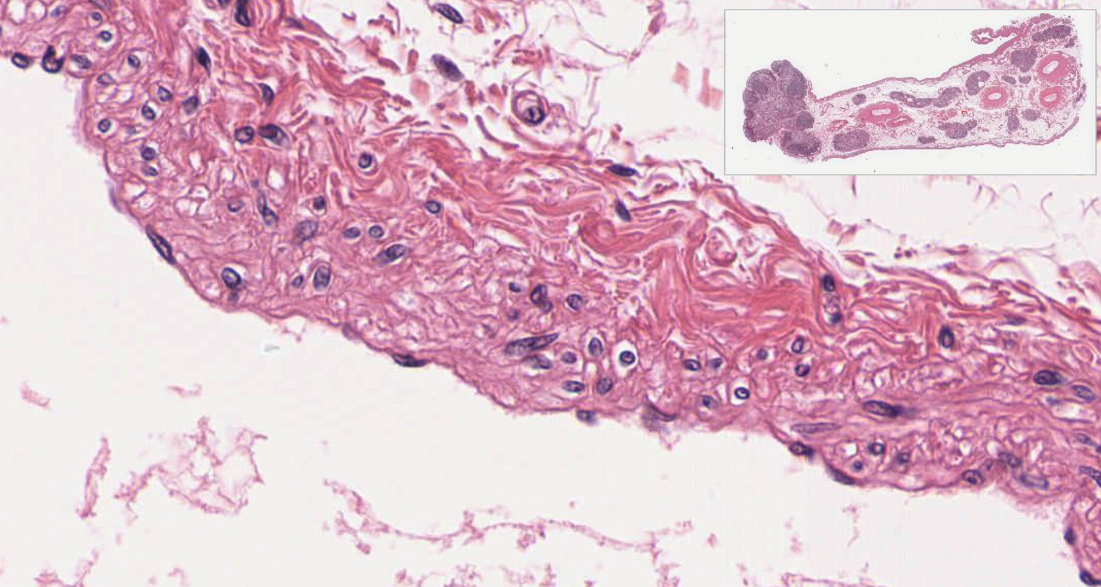
The epithelial tissue is a type of body tissue that forms the covering on all internal and external surfaces of your body, lines body cavities and hollow organs and is the major tissue in glands. Epithelial tissue has a variety of functions depending on where it’s located in your body, including protection, secretion and absorption.
Epithelial Tissue in the Heart
The epicardium is a layer of epithelial cells covering the surface of the heart. The epicardium migrates to the surface of the heart from villous projections from the septum transversum in mammals. The epicardium serves as a progenitor source, contributing multipotent cells that give rise to cardiac mesenchyme.
Epithelial tissue in Veins
Epithelial Tissue in Arteries
Epithelial Tissue in Capillaries
Connective Tissue
How does the Cardiovascular system responds to change?
The Cardiovascular System has many ways to maintain homeostasis. Some homeostatic functions of the cardiovascular system is the transport of nutrients and wastes, defending the body against invaders, and distributing heat to regulate body temperature. Also, the heart pumps harder/softer depending on stress, anxiety thickness of blood, diameter of vein, velocity of vein, caffeine intake, and alcohol intake. For example, when you are hot your blood rushes to skin to make you sweat to cool you off. Also, when your blood is thicker your heart has to beat harder to get the blood to flow through your body.

How does the Cardiovascular System respond to change in pH?
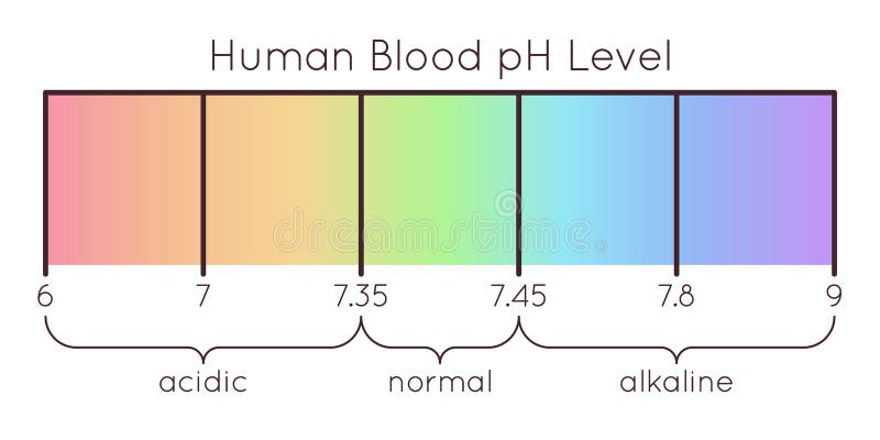
When the blood pH decreases, it indicates that the carbon dioxide level is increased in the bloodstream. Therefore, the heart sends deoxygenated blood to the lungs in more quantity to stabilize the carbon dioxide level in the body. As a result, the heart rate increases. However, when pH increases the opposite occurs. When the pH of the blood increases, the heart rate decreases. In order to restore the heart rate, the deoxygenated blood is delivered to the lungs at a maximum rate to remove the carbon dioxide.
How does the Cardiovascular System respond to change in blood pressure?
Blood pressure is regulated in the body by changes to the diameters of blood vessels in response to changes in the cardiac output and stroke volume. Factors such as stress, nutrition, drugs, exercise, or disease can invoke changes in the diameters of the blood vessels, altering blood pressure.
How does the Cardiovascular System respond to change in blood sugar?
The circulatory system allows blood glucose levels to be regulated. The hormone glucagon, carried in the blood, signals the liver to release glucose into the blood and the presence of insulin in the blood instructs the cells to take in glucose from the blood.
